Hallux Rigidus

fb_img_1474919252840


Hallux Rigidus


Fall is upon us here in Anchorage.  We hope that you, like us here Alliance Foot and Ankle,  have lingering beautiful memories of clear skies, great evenings with friends and family,  beautiful mountain vistas, and fish in your freezer. Or maybe your most lingering part of summer is foot pain.  If so, we are here to help.

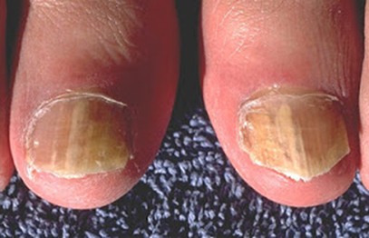
At our practice, we notice that once the weather starts to cool and the days become shorter and our patients find them in our office rather than on the mountain side. One of the more common problems we see with our very active community is pain in the joint of the big toe called hallux rigidus.  Common symptoms are pain deep within the joint, a large spur on the top of the joint and decreased range of motion.

This problem usually occurs very gradually. Perhaps you have had occasion discomfort for years or you are just starting to notice a painful bump on your great toe joint.  For many people, this can cause significant decrease in doing the recreational activities they love or even difficulty on the job.


What causes it?

There are usually two common causes of hallux rigidus. The first cause is chronic repetitive trauma to the joint that causes a jamming motion such as running, climbing or stubbing your toe can damage the cartilage in the joint. Over time the cartilage may start to erode and your body lays down more bone to protect itself which is what causes the bone spur you may feel at the top or side of the joint.

hallux-rigidus-diagram

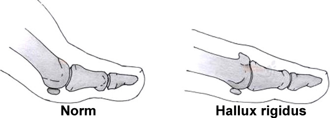
The second cause is a biomechanical problem related to how the bones in the great toe joint act. The two bones that involved are the first metatarsal (the long narrow bones of the midfoot just behind the toes) and the proximal phalanx (the bone at the base of your great toe). These two bones should line up well allowing the toe to easily glide over the smooth cartilage of the 1st metatarsal. Often times people are born with a slightly more elevated 1st metatarsal. This causes jamming when the great toe attempts to move up and over the head of the metatarsal bone. Over time, this chronic jamming leads to wearing away of the cartilage and extra bone being deposited.

hallux-rigidus-b


How can it be treated?

Fortunately, we have a variety of ways to treat this condition both from a conservative and surgical approach.  The more conservative (non-surgical) approaches involve orthotics that you wear in your shoe to restrict motion of the joint which helps alleviate some of the pain. Icing and anti-inflammatory medications such as Ibuprofen or Naproxen can provide temporary help.  Cortisone injections can also be of great benefit in reducing the swelling and discomfort.

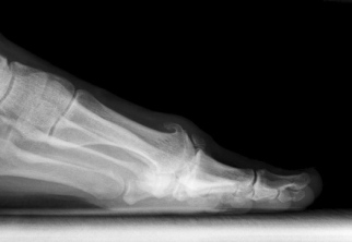
For some people, conservative measures simply aren’t enough. We offer a variety of surgical procedures to correct the issue and return you to your activities.  During an office visit, x-rays are taken and reviewed with you by one of our doctors to determine if a surgical procedure is a good fit for you.  The procedure chosen is based on the severity of the damage done to the joint, the underlying cause of the hallux rigidus, and the patient’s goals after surgery. Possible procedures are cheilectomy which involves shaving down the painful prominent bone, bone cuts to get correct the alignment of the joint, joint replacement or joint fusion (arthrodesis).

If you think you suffer from hallux rigidus or any other foot problem, we would be happy to help you get back on your feet again.


Dr. Kristin Klingenstein DPM

 fb_img_1474996452504


High Arched Foot Treatment in Anchorage, AK

Untitled-23

The cavus, or high arched foot, is described as having a high medial longitudinal arch. The shape is caused by the osseous structure of the hindfoot and forefoot. A patient may have a reducible (supple) nature. There are many etiologies for a cavus foot type. Many stem from an inherited neuromuscular disorder. Not all high arched feet are symptomatic. In some, symptoms may occur in the foot, ankle, or leg. Claw toes (flexion at the proximal and distal interphalangeal joints) are very common with cavus foot types. Painful callosities may develop on the dorsal aspects of the digits and the plantar aspects of the metatarsal heads. With hindfoot or combined cavus, a prominent posterior aspect of the calcaneus may occur, which can be irritated by the heel counter or the shoes. Severe cramps in the legs may result from a true or pseudo Achilles tendon equinus (inability to lift the foot up at the ankle). In conditions caused by peripheral nerve pathology (i.e., Charcot Marie Tooth disease) tendon imbalances may occur, including the loss of entire muscle groups which can lead to a drop foot condition.

Proper diagnosis is needed to assess whether neurologic pathology is present and, if so, to determine its classification and stage. This is important to determine if the condition may progress overtime.

The cavus (high arch) is a clinical challenge, especially in progressive conditions. Many times conservative treatments provide pain-free ambulation. Treatment is initially aimed at alleviating painful callosities by proper shoeing, accommodative orthoses, or an AFO (ankle foot orthoses) if a drop foot is present. If conservative and palliative measures fail to alleviate symptoms, surgical reconstruction is considered. Using a combination of soft tissue(i.e., tendon transfers, releases) and osseous procedures, the painful cavus foot is reconstructed. For some cavus conditions, digital deformities (single or multiple) are all that need correction, but for others, multiple hindfoot fusions with tendon transfers from the posterior leg are necessary (i.e., to treat a painful hindfoot with a drop foot).

Visit http://alaskapodiatry.com/ for more information.

Plantar Wart Treatment in Anchorage, AK

Untitled-22

Warts (Verruca Plantaris) 

Plantar warts (Verrucae plantaris) are a common problem affecting the feet.  They are caused by a viral infection within the skin.  The virus enters the skin directly and is typically transmitted by close contact.  Swimming pools, bathrooms, and locker rooms are common areas associated with the spread of plantar warts.


Warts reach their peak incidence between the ages of 12 and 16, then decline in frequency.  Many treatments are available and they all share one characteristic: there is a 10% recurrence rate. Although the actual mechanism is unknown, approximately 30 % of warts may spontaneously disappear within a period of three though six months.  On the other hand, without treatment warts may persist for years, often increasing in size and number.  Because warts are contagious they are infectious not only for the individual, but for the family and community as well.

Pain is typically felt clinically from side to side palpation. Upon debridement, multiple pinpoint bleeding sites are observed which are pathognomonic. This condition is called papillomatosis and results from cutting the ends of multiple capillaries that have proliferated from within the papillary layer of the dermis into the verruca.

Prevention of plantar warts is much enhanced by careful hygiene and the use of personal sandals or aqua socks when utilizing facilities that me a source of the virus.  Walking without shoes is often the initiating event of this problem, as well as many other foot disorders and injuries.  Wearing shoes without socks or stockings can result in reinfection.

This common, yet bothersome and persistent problem can be well controlled by professional treatment.  The treatment that your physician recommends will be based upon your age, occupation and expectations.  Topical acid creams or plasters combined with debridement are the main forms or treatment. Occlusal (salicylic acid in a flexible base) works very well and has high patient acceptance. Blunt enucleation with application of phenol to or electrical desiccation of the base of the verruca is a quick definitive approach that can be used as a primary or secondary treatment. The use of Vitamin A and Zinc has been beneficial in treating warts and is also useful to prevent recurrence.  These supplements are present in many multi-vitamin preparations or they can be purchased separately and are found in the nutrition section of many large grocery stores.

Peripheral Neuropathy Treatment in Anchorage, AK

Untitled-21

Peripheral neuropathy refers to disorders of the nerves which are outside of the central nervous system (nerves which are away from the brain and spinal cord).  Unfortunately, this problem is all too common.  Peripheral neuropathy can have various causes and produce many different symptoms.

Symptoms that involve the feet may include burning, tingling, numbness, or shooting pains.  Numbness often affects the bottom of the toes and balls of the feet.  Peripheral Neuropathy affects the longest nerves in the body first, which are the nerves that start from the bottom of the spinal cord and extend to the toes.  It is not uncommon for the big toe to be involved first.  If symptoms progress, the hands can become involved as well.  Some experience a sensation as if a band was around the foot or that a stocking was on when it is not.   At times the feet may be overly sensitive.  Even common sensations can be irritating such as the wearing of shoes, walking on carpet, etc.

Causes of Neuropathy

There are many causes of peripheral neuropathy.   It is quite common, however, that an exact cause cannot be discovered.  Poor circulation is usually not the cause of numbness.  So, numbness does not necessarily mean you are at risk as a result of poor blood flow!  Your health care provider will check your circulation to be sure it is adequate.

Known causes of peripheral neuropathy include:

¨ Diabetes (the most common cause)

¨ Alcohol toxicity

¨ Thyroid abnormalities.

¨  Nutritional deficiencies such as inadequate Vitamin B-12 or the  inability of the body to utilize vitamins and nutrients.

¨ Illnesses such as Guillian-Barre.

¨ Inherited conditions that affect nerves.

¨ Entrapment of nerves can occur with back problems, sciatica, or  tarsal tunnel (a pinched nerve near the inside of the ankle similar to carpal tunnel).

¨ Growths that press on nerves.

¨ Injury to nerves.

Drugs used in the treatment of cancer, HIV/AIDS, and other conditions

Treatment of Neuropathy

Treatment for peripheral neuropathy depends on the cause.  Blood tests or nerve conduction studies may be used to check for various causes.  If an underlying cause can be identified, such as diabetes, then treating that cause is important.  Injections or even surgery in some cases can relieve a pinched nerve.

Though symptoms may sometimes improve, the sensations such as numbness do not always go away.  If pain is associated with neuropathy, then your health care provider may recommend trying a cream such as capsaicin which is available without a prescription.   Your health care provider may also recommend the use of oral medications to treat neuropathic pain.  The types of medication often used to treat neuropathy are for other conditions such as seizures or depression.  Common medications that may be prescribed are Neurontin, Lyrica, or Cymbalta to name a few.  Many individuals find that neuropathy is most irritating or painful when trying to go to sleep as there are no distractions from the irritation.  Occasionally, neuropathy medications are combined with pain medications, such as Ultram, to help patients through the initial period as the Neuropathy medications may take some time to reach their maximum benefit.  The important point to remember is there is treatment available, which is tailored to fit each individual patient.

Visit http://alaskapodiatry.com/ for more information.

Metatarsalgia Treatment in Anchorage, AK

Untitled-20

Metatarsalgia is a condition that presents with pain to the front of the foot (ball or forefoot) that usually results from activity and/or faulty foot mechanics.

A review of anatomy is necessary to explain this common condition. The toe bones (phalanges) two in the big toe, three in toes two through five, and the metatarsal bones (long bones in ball of foot), meet at the ball of the forefoot where the toes flex up and down to form the metatarsal- phalangeal joints. These joints are numbered one to five from big to little toe. When standing the metatarsals have a downward slope and we stand on the front part of the bones called the heads. All five metatarsal heads should bear an equal amount of weight, one-fifth or 20% of the forefoot load to each metatarsal head. The space between each metatarsal is called the intermetatarsal space. Among the anatomy that resides in these spaces are structures called bursas. A bursa is a fluid filled cavity or balloon type structure that acts to provide cushion and shock absorption.

The intermetatarsal bursa are found between metatarsal heads two though five and are small, about the size of a little grape. As we walk or run a tremendous amount of weight or load is presented to the forefoot. With proper foot function and anatomy the forefoot load is absorbed and pain free ambulation is achieved. Improper function or faulty anatomy can result in activity induced pain to the ball of the foot known as metatarsalgia. This term is non-specific. It does not define the reason why a person is experiencing pain to the forefoot. A thorough history and physical exam along with possible x-ray studies is necessary to determine the cause of a persons metatarsalgia symptoms.


Anatomical variations can lead to metatarsalgia symptoms. A person may have a metatarsal that is longer or shorter than the adjacent bones. A metartarsal bone can be positioned too high or low in comparison to the other metatarsals. With these variations all five metatarsal heads may not be on the ground equally, thus not bearing their 20% of the forefoot weight load. Anatomy of a metatarsal can be altered by trauma, such as a break/fracture, thus changing the position and function of that bone. This may result in more or less pressure to the effected bone, or the adjacent bones. Arthritis (degenerative joint disease) can lead to painful function at the metatarsal- phalangeal joints. A tight heal cord/Achilles tendon (a condition called equines) can lead to increased pressure to the forefoot. A high or low arch can lead to altered pressure across the metatarsal heads. Neuromuscular conditions can alter the way a person walks/runs resulting in uneven weight distribution to the forefoot.

Improper or faulty foot mechanics may lead to metatarsalgia symptoms. If there is uneven weight distribution across the ball of the foot, swelling to the bursa between the metatarsal heads may occur. Many people describe this as if their socks are bunching up under the ball of the foot or they are walking on a lump. If a bursa remains swollen it can pinch the adjacent intermetatarsal nerve (which is a nerve headed towards the tips of the toes to give sensation around the nail) and lead to a neuroma. A neuroma is a swollen nerve in the intermetarsal space that can present with sharp, electrical type symptoms that can radiate towards the toes becoming increasingly debilitating.

Once the cause of an individuals metatarsalgia is identified a treatment protocol can be established. Most treatment is non-surgical. If faulty foot function is contributing, adding an orthotic (shoe inlay) along with the use of proper supportive shoes may be very helpful. Many different types of orthotics are available depending on the nature of the problem. Self directed or formal physical therapy can help with tight or weak muscles and tendons, muscle group imbalance, and inflammation (swelling) involving soft tissue structures (bursa), joints, and irritated nerves. Oral (medication) and injectable anti-inflammatory medication can have a dramatic effect on metatarsalgia symptoms. Many times a combination of conservative treatments are necessary to achieve pain free ambulation. Surgical treatments are reserved for conditions requiring that something needs to be fixed, such as a fractured metatarsal bone that has not healed or healed improperly, or a metatarsal bone that is dramatically out of position. A chronically swollen, painful neuroma that has been unresponsive to conservative care may respond well to surgical options also.

In most cases once the diagnosis of metatarsalgia is made and the likely cause indentified, non-surgical treatment protocols alleviate most patients symptoms.

Visit http://alaskapodiatry.com/ for more information.

Hammer Toe Treatment in Anchorage, AK

Untitled-19

Thanks to Randy for posting a question, hope this helps.

A hammertoe is a contracture—or bending—of one or both joints of the second, third, fourth, or fifth (little) toes. This abnormal bending can put pressure on the toe when wearing shoes, causing problems to develop. Common symptoms of hammertoes include:

• Pain or irritation of the affected toe when wearing shoes.

• Corns (a buildup of callus skin) on the top, side, or end of the toe, or between two toes. Corns are caused by constant friction against the shoe. They may be soft or hard, depending upon their location.

• Calluses (another type of skin buildup) on the bottom of the toe or on the ball of the foot.

Corns and calluses can be painful and make it difficult to find a comfortable shoe. Even without corns and calluses, hammertoes can cause pain because the joint itself may become dislocated or arthritic.

Hammertoes usually start out as mild deformities and get progressively worse over time. In the earlier stages, hammertoes are flexible and the symptoms can often be managed with noninvasive measures. But if left untreated, hammertoes can become more rigid and will not respond to non-surgical treatment. Corns are more likely to develop as time goes on—and corns never really go away, even after trimming. In more severe cases of hammertoe, open sores may form.  Because of the progressive nature of hammertoes, they should receive early attention.

Causes of Hammertoes 

The most common cause of hammertoe is a muscle/tendon imbalance. This imbalance, which leads to a bending of the toe, results from mechanical (structural) changes in the foot that occur over time in some people.  Hammertoes are often aggravated by shoes that don’t fit properly—for example, shoes that crowd the toes. In some cases, ill-fitting shoes can actually cause the contracture that defines hammertoe. For example, a hammertoe may develop if a toe is too long and is forced into a cramped position when a tight shoe is worn. Occasionally, hammertoe is caused by some kind of trauma, such as a previously broken toe.

Treatment:

Non-Surgical Approaches

There are a variety of treatment options for hammertoe. The treatment your  foot and ankle surgeon selects will depend upon the severity of your hammertoe and other factors.

A number of non-surgical measures can be undertaken:

  • Trimming and padding corns and calluses.
  • Changes in shoewear. Avoid shoes with pointed toes, shoes that are too short, or shoes with high heels—conditions that can force your toe against the front of the shoe.
  • Orthotic devices. A custom or OTC orthotic device placed in your shoe may help control the muscle/tendon imbalance.
  • Medications

When Is Surgery Needed?

In some cases, often when the hammertoe has become more rigid, surgery is needed to relieve the pain and discomfort caused by the deformity.  Your surgeon will discuss the options and select a plan tailored to your needs.  Among other concerns, he or she will take into consideration the type of shoes you want to wear, the number of toes involved, your activity level, your age, and the severity of the hammertoe.

One type of surgical procedure performed to correct a hammertoe is called arthroplasty. In this procedure, the surgeon removes a small section of the bone from the affected joint.  Another surgical option is an arthrodesis.  Arthrodesis is a procedure that involves fusing a small joint in the toe to straighten it. A pin or other small fixation device is typically used to hold the toe in position while the bones are healing.

It is possible that a patient may require other procedures, as well— especially when the hammertoe condition is severe. Some of these procedures include tendon/muscle rebalancing or lengthening, small tendon transfers, or relocation of surrounding joints.  Often patients with hammertoes have bunions or other foot deformities corrected at the same time. The length of the recovery period will vary, depending on the procedure or procedures performed.

Visit http://alaskapodiatry.com/ for more information.


Hallux Rigidus/Limitus Treatment in Anchorage, AK


Untitled-18

Hallux Limitus/ Rigidus is a condition where the big toe joint degenerates and becomes stiff and arthritic.  The hallux (big toe) needs to flex upward and downward at the first metatarsal phalangeal joint (big toe joint) to function normal.  Due to certain conditions such as trauma/ injury, genetic predisposition to faulty structure, generalized (whole body) arthritic conditions or prior surgery, the big toe joint may start a process known as degenerative joint disease.  This progressive condition leads to wearing out of the articular cartilage (the pearl white shinny tissue seen where two bones form a joint), and the big toe joint starts to become stiff and painful to move.  Once the body recognizes that this process is occurring extra bone is laid down around the joint (bone spur) to begin a process of fusing the joint solid thus no motion, no pain.  While this process is occurring pieces of the extra bone can break off into smaller fragments and cause pain to the joint.


Hallux limitus is more common in men than women but plenty of active women are diagnosed with the condition.  It consists of four stages.  Most people are unaware of any symptoms in stage one other than possible progressive joint stiffness.  Many present for treatment in stage two as the joint becomes more painful to move and fracture fragments may start to jam the joint.  If left untreated as one progresses toward stage four the joint becomes very stiff and bulbous as the body places bone around the joint in an attempt to fuse it (no motion, no pain).  Small amounts of motion though stiff occurs and the poor quality of this motion combined with extensive bone spur development (some pieces may even be fractured) can lead to extreme pain with ambulation.


Treatment is patient specific depending on age, activity level, type of employment, stage of condition, prior treatment, shoe choices, and expectations.  Hallux limitus is progressive over time and is not reversible.  Non-surgical treatments may alleviate pain and slow progression.  Stiff sole shoes with an appropriate orthotic (shoe inlay) may off load the great toe joint and support the transverse and longitudinal arch of the foot.  Anti-inflammatory medications and injections into the joint can temporarily ease symptoms.  Self-directedor formal physical therapy may help improve function, decrease swelling, and increase local blood flow.  As the condition progresses with increased pain and decreased function, surgical options are usually considered.  If it is possible, an attempt is made to reconstruct the degenerative joint to reduce the stage of the condition.  This preserves ones natural joint and although far from perfect may function pain free for many more years.  As one advances to stage three or four, reconstructing ones joint may not be possible and joint destructive procedures such as a joint implants or surgical fusion need to be considered.  Joint implants may be limited to a select type of person.  Joint fusion (arthrodesis) is a very powerful procedure to eliminate pain and still allow for a high level of activity.  Joint fusion requires a committed rehabilitation and understanding that adjacent joints can be affected in the future.

Visit http://alaskapodiatry.com/ for more information.


Gout Treatment in Anchorage, AK

Image

Gout is caused by an elevation of uric acid within your blood.  Uric acid is a natural substance in your body that is a breakdown product of proteins.  Gout occurs in people who either make too much uric acid, eat too many foods that are high in uric acid, or in people who cannot get rid of excess uric acid in their blood.  Gout medicines focus on helping your body produce less uric acid, or helping your body eliminate excess uric acid.

Where does Uric Acid come from?

You naturally produce most of the uric acid in your body, but there is a percentage (up to 20%) of uric acid that comes into your body through diet.  Adjusting your diet by avoiding foods high in uric acid can help reduce your chances of repeat gout attacks.  In addition, keeping yourself very well hydrated with water is helpful.  Some medications can make a gout attack more likely, including some blood-pressure pills (water pills).  Your doctor may adjust these medications if gout becomes a problem.

In addition to medicine, you can help avoid future gout attacks by following the following diet guidelines:

Eliminate the following:

Liver    Kidney     Anchovies      Sweetbreads   Sardines      Brains     Heavy gravies      Heart    Fish roes    Mussels     Herring    Beer / Wine    Meat extracts, consommé


Reduce the following foods to no more than one serving per day:

Meats     Beans   Fowl     Peas   Cauliflower   Mushrooms Other seafood   Lentils     Spinach Yeast      Asparagus    Whole grain cereals    Fish (except noted above)

These foods do not cause gout:

Fruits     Milk       Cheese    Eggs     Refined cereals   Nuts   Spices (including salt)   Sugars & Sweets    Vegetable soups (clear)    Vegetables


Diagnosis & Treatment

A blood test may be used to test the uric acid level.  It is common; however, that the blood level of uric acid may be normal even while a person is having a gout attack.  If the diagnosis is in question, a sample of joint fluid may be taken and tested for gout crystals.  Most cases of gout affect the big toe joint but other areas can also be affected.  The pain can be quite severe and result in joint swelling and redness.  Walking may be difficult and pressure of a shoe or even bed covers can be painful.  Treatment for the acute, severe pain may include anti–inflammatory medications (such as indomethacin or colchicine) or injections near the area.  A sandal or surgical shoe may be useful to relieve pain as well.

Visit http://alaskapodiatry.com/ for more information.

Ganglion Cyst Treatment in Anchorage, AK

ganglion

What is a ganglion?       

Ganglion Cysts are a benign fluid-filled lump (cyst) that can occur in many areas of the body, but is most common on the hands and feet.  They typically arise over a joint or tendon.  Although the exact causes are not known, a ganglion likely occurs due to a weakness in the covering of the tendon or joint space. Direct injury or everyday minor trauma can potentially be a cause.  Motions of a tendon over a bony prominence or pressure from a shoe on a tendon or joint are examples of minor trauma.  Weakness in the covering with a subsequent swelling of liquid into a confined space causes the lump to form.  The growth is usually very firm but may be somewhat soft, depending on the fluid content.  The fluid itself is usually similar in appearance to a thick clear jelly.

Diagnosis

A diagnosis of ganglion cyst is usually made on clinical examination based on location of the growth and pertinent history.  An x-ray may be taken to rule out a bone spur as the aggravating factor behind the formation of the cyst.  If there is any question as to the extent of the growth, an MRI may be ordered for a more detailed evaluation.

Treatment 

Proper treatment is based on pain, size, location and aggravating factors. Once a mass is diagnosed as a ganglion cyst it may be left alone, untreated, if it is not painful or causing problems with shoes. Occasionally these growths will spontaneously disappear.  Cysts can be drained by needle aspiration followed by a cortisone injection in an effort to prevent the cyst from filling again.  Once drained, a compression dressing is worn over the site for a period of time in an attempt to reduce recurrence.  Over half of the cysts will refill with this form of treatment, but the upside is that the procedure can be repeated again if needed.  If the growth is a source of pain or irritation and drawing fluid out (aspiration) has failed, then surgical excision may be necessary. Although surgical excision reduces the chance for recurrence ganglions will sometimes come back.  Surgical excision is an outpatient procedure.   Crutches and non-weight bearing are not usually needed.   A surgical shoe is usually worn for 2 to 3 weeks during the time that the incision is healing.  There really is no preventative care for ganglion cysts due to their spontaneous nature.

It is important to be evaluated by a foot and ankle specialist to ensure proper diagnosis.

Visit http://alaskapodiatry.com/ for more information.

Fungus Infection Treatment in Anchorage, AK

Image

Fungal infection of the foot, or Tinea pedis (athletes foot), is a superficial infection caused by a dermatophyte. Dermatophytes are fungi that thrive in a nonviable tissue of the skin, nails, or hair. Trichophyton and Epidermophyton are the principle genera causing infection. Dermatophytosis can involve the entire plantar foot with extensive scaling, hyperkeratosis or even bulla formation. Infection between the toes (Tinea pedis interdigitalis) is quite common. When involvement of the nails occurs (onychomycosis) they take on a very thick, discolored (yellow/brown) appearance. Diagnosis is usually clinical, but a fungal culture or potassium hydroxide mount (KOH) of skin or nail scrapings can help diagnose unclear presentations by showing the presence of a fungal organism within the sample.

 Tinea pedis can usually be resolved after several weeks of treatment with topical medications. Oral antifungals (i.e., Lamisil, Griseofulvin,) can be used for extensive infection or poor topical response. Onychomycosis is more difficult to eradicate. Topical therapy results are poor, and recurrence is common after completion of successful oral treatment. Temporary nail avulsion or permanent nail removal (matrixectomy) may be indicated for several deformed, painful onychomycotic nails.

Visit http://alaskapodiatry.com/ for more information.